Skin Cancers & Treatments
Skin cancer is the most commonly diagnosed cancer in Australia, with approximately two in three Australians diagnosed with some form of the disease before the age of 70. Unenviably, we have one of the highest rates of skin cancer in the world, including Basal Cell Carcinoma, Squamous Cell Carcinoma, and Melanoma. This is two to three times the rate seen in Canada, the US, or the UK. The majority of skin cancers are caused by over-exposure to ultraviolet radiation from the sun.
Other risk factors include age, a fair skin type, the use of solariums, previous radiation therapy, occupational exposure to certain chemicals, and in some types of skin cancer, family history is also relevant. Whilst certain people may be more at risk, the simple message is that anyone, of any age, gender, or ethnicity can develop skin cancer and that as a patient you can play a role in the prevention and treatment of this disease by:
Routinely wearing sunscreen and engaging in sun-protective behaviours.
Performing regular self-skin examinations - keeping an eye on changing skin patterns or spots on your own skin is a great first defence.
Seeing your GP or Dermatologist for a full skin examination when recommended or when you are concerned regarding a new or evolving lesion.
Phone The Skin Centre on (07) 5597 7170 to make an appointment to see your Dermatologist or email reception@skincentre.com.au
Benign, Precancerous and Cancerous Skin Lesions
The full skin examination may reveal a number of different benign, premalignant or malignant skin lesions. These may arise from any structure of the skin including the epidermis and dermis, the hair follicle, glandular structures, blood vessels and nerves. The Dermatologists we support have attempted to provide a description of the more common skin lesions seen in their practice, and in Australia, but by no means does this list reflect the vast array of skin tumours and cancers that we treat. If you can’t find the information that you need, and wish to make an appointment to see your Dermatologist – please call The Skin Centre on ph: 07 5597 7170.
The term benign refers to something which is harmless. Some of the more common benign skin lesions include seborrhoeic keratoses, acrochordons, sebaceous hyperplasia and cysts.
Premalignant refers to a situation in which a collection of skin cells has become abnormal but is yet to become cancerous. Despite being pre-cancerous, your Dermatologist may still suggest treatment to prevent their transformation into something more sinister. Of the various pre-malignant lesions, one of the most commonly encountered is the Actinic Keratosis, which you can read about below.
Malignant skin cancers are those where the cells have now become abnormal and have gained the ability to invade nearby tissue. Malignant cells can spread to distant sites so treatment for these forms of skin cancer is recommended. Non-melanoma skin cancers, such as intra-epidermal carcinoma (IEC), squamous cell carcinoma (SCC), basal cell carcinoma (BCC), and melanoma are all considered malignant and information about them can be found below. Certain lesions can also be considered markers for an increased risk of other cancers, such as Dysplastic Naevi (an atypical mole).


Premalignant Non Melanoma Skin Cancers (Premalignant NMSC)
Actinic Keratosis (AK) (Solar Keratosis)
Actinic Keratoses (AK) are premalignant skin lesions seen often in Australia. They are scaly, red and thin plaques that appear on the areas of the skin that are frequently exposed to the sun. They tend to be more common in fairer individuals, outdoor workers, men and older patients. They feel rough to the touch and people may repeatedly pick and scratch at them, mostly unintentionally. Their risk of transforming into a cancerous lesion, for example an intra-epidermal carcinoma (IEC/Bowen’s Disease) or Squamous Cell Carcinoma (SCC) ranges from between 0.075%-0.096% per year. Whilst it isn’t strictly necessary, treatment of these lesions is often requested by the patient, who finds their appearance unsightly. Otherwise it may be initiated by the doctor as part of a field treatment of an area of skin that is marked by sun spots. Most people are familiar with their family doctor treating these areas with cryotherapy but other options include curette and cautery (C&C), photodynamic therapy (PDT), chemical peels, laser ablation or topical treatments including 5-Flurouracil (Efudix), imiquimod (Aldara) and topical diclofenac (Solaraze).
Malignant Non Melanoma Skin Cancers (Malignant NMSC)
Intra-Epidermal Carcinoma (IEC) (Bowen’s Disease/Squamous Cell Carcinoma in Situ)
An intra-epidermal carcinoma (IEC) is a superficial skin cancer that can appear unexpectedly or may originate from a pre-existing actinic keratosis. An IEC appears most commonly as a slow-growing, persistent red scaly patch in sun exposed sites. People at increased risk of this skin cancer include older patients, fairer skin types, people whose immune systems are impaired by illness or medication. Variants of these lesions are seen in certain sites of the body, including genital Bowenoid Papulosis or Erythroplasia of Queyrat. Depending on the type, some of these lesions may have a higher risk of progressing to Squamous Cell Carcinoma. Treatment to remove these malignant lesions is by either curette and cautery (C&C), topical treatments, photodynamic therapy (PDT), wide local excision (WLE) or Mohs micrographic surgery (MMS).
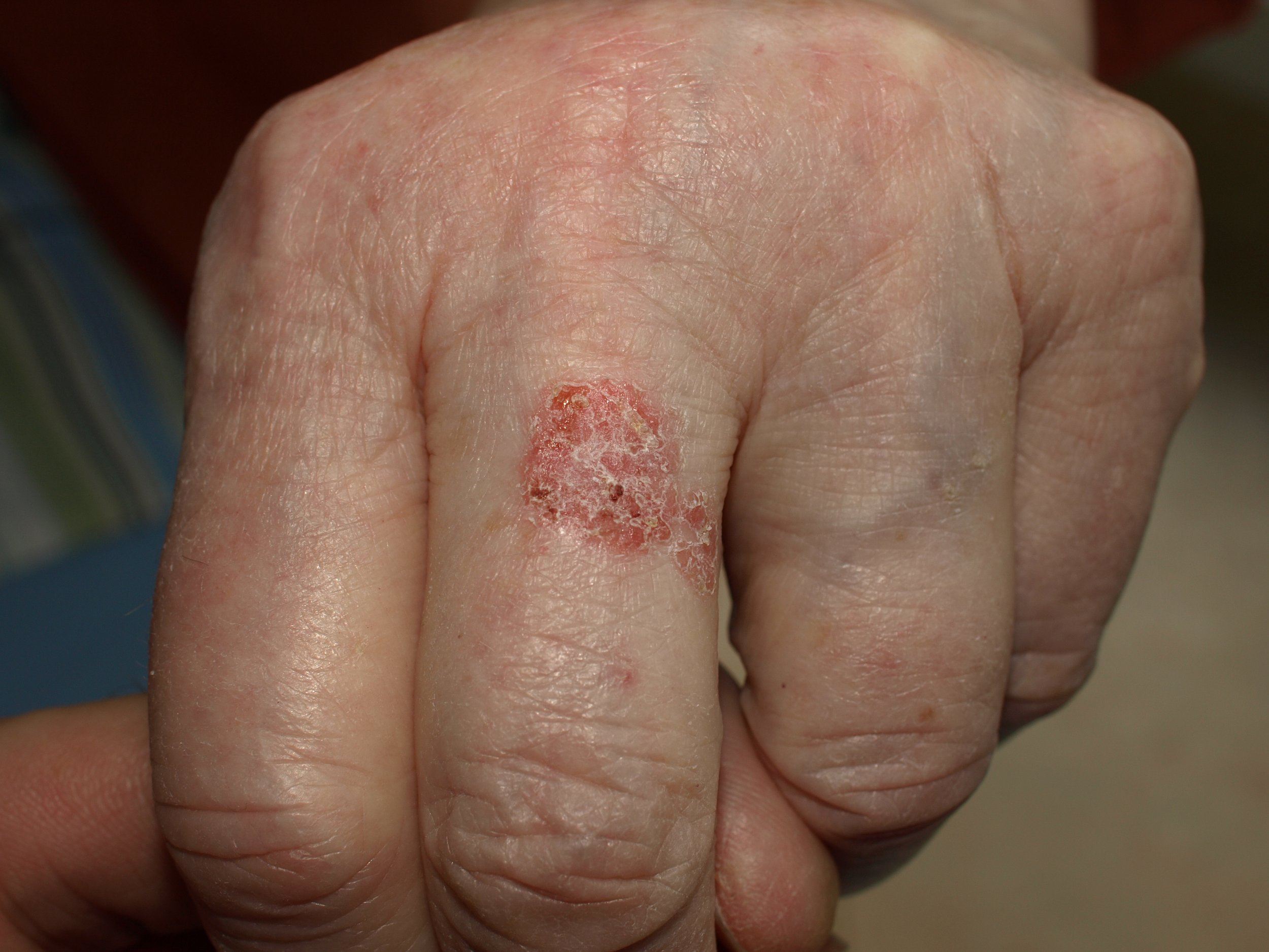
Squamous Cell Carcinoma (SCC)
A Squamous Cell Carcinoma (SCC) is an invasive form of skin cancer that accounts for 30% of all non-melanoma skin cancers in Australia. It can be diverse in its appearance, ranging from a lesion that is flat, red and scaly to a nodule that can bleed and crust over. SCC can sometimes appear as a non healing sore or a lesion that is painful to touch. Although they can be found anywhere on the body, they tend to favour the head, neck, forearms, hands and legs. Once one appears, it will generally grow quite quickly, evolving over weeks to months. There are many factors that increase a person’s risk of developing SCC including chronic sun exposure, a history of previous skin cancer, older age, fair skin, radiation therapy or chemical exposure, certain genetic syndromes and immunosuppression (organ transplant, blood cancer, medication). A diagnosis of SCC is made on biopsy, but further investigations may be undertaken if your Dermatologist is concerned this cancer may have spread to other parts of the body. Treatment options depend on the subtype, the site and other patient factors but may include curette and cautery (C&C), wide local excision (WLE), Mohs micrographic surgery (MMS) and in some cases radiation treatment (this may be combined with surgery where the cancer involves a nerve). Whilst SCC is an invasive skin cancer, if detected and treated early, the prognosis is generally good.
Keratoacanthoma (KA)
A Keratoacanthoma (KA) is considered a special variant of a well differentiated squamous cell carcinoma (SCC), which unlike other forms of SCC, may resolve without treatment. Often described as a little volcano, it appears suddenly and is frequently mistaken as a painful pimple with a central crater containing a crusty core. They generally appear as a single lesion, but can also occur in groups or clusters. They are most commonly found on the sun exposed areas of the skin such as the head, neck, arms and legs. Other than sun damage, certain medications and immunosuppression can all increase a patient’s risk of developing a KA. Although a KA may disappear on its own, it is often removed by biopsy for diagnosis under the microscope. As it can be difficult to distinguish these lesions both clinically and histologically from more aggressive forms of SCC, removal is sometimes recommended and can be achieved through curette and cautery (C&C), wide local excision (WLE) or Mohs micrographic surgery (MMS).
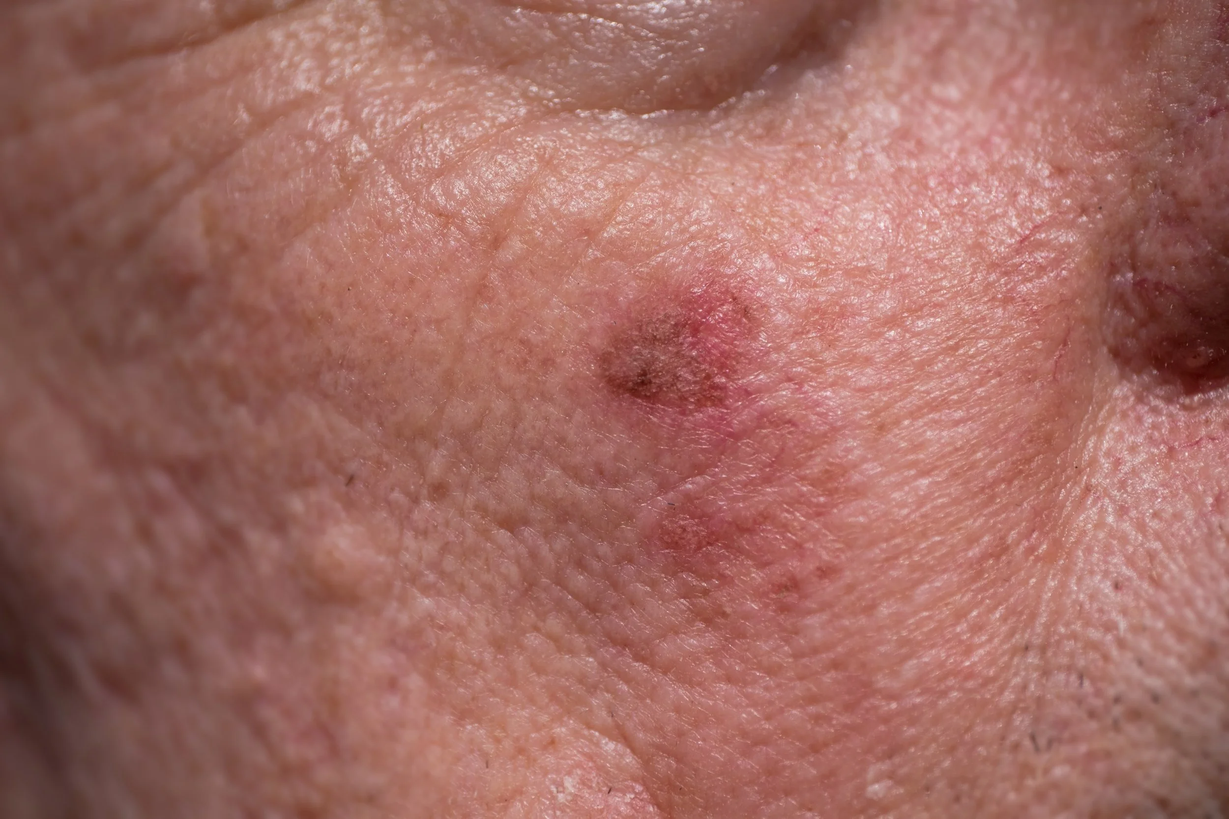
Basal Cell Carcinoma (BCC)
A Basal Cell Carcinoma (BCC) is the most common form of skin cancer accounting for 70% of all non-melanoma skin cancers in Australia. Fortunately, they are slow growing cancers that virtually never spread to other parts of the body and with early detection and treatment are usually completely curable. However, they can grow quite large, destroying surrounding structures, therefore removal when they are small is best to avoid extensive surgery. There are several forms of BCC with a diverse range of clinical features. They can appear as slow growing thin plaques, pink or pearly nodules, a non healing sore or a scar like area of skin depending on the subtype. Sometimes the edge of the BCC can be difficult to detect and the skin cancer may in fact be much larger than it appears. Diagnosis of these lesions is often delayed as the may indistinct in their early stages. The sun plays the most important role in the development of BCC, however fair skin, a family history and certain genetic syndromes can all predispose to increased risk of disease. After diagnosis has been made on biopsy, there are several treatment options available for BCC depending on the size, type, location of the lesion and patient factors such as preference, age and health. These treatment options include topical agents, cryotherapy, curette and cautery, photo-dynamic therapy, wide local excision, Mohs micrographic surgery, radiotherapy and in some cases oral chemotherapy.
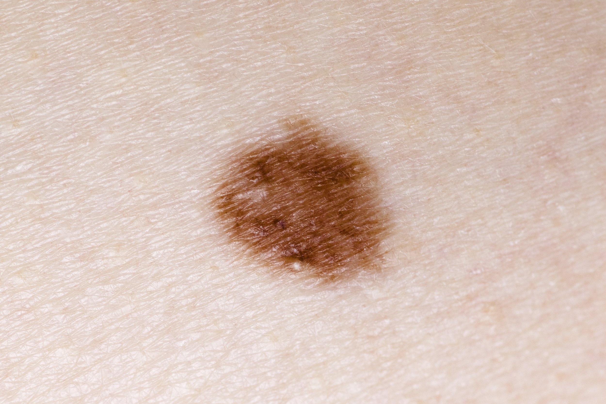
Dysplastic Naevi (Atypical Melanocytic Naevi)
A naevus is a benign growth occurring on the skin, better known as a ‘mole’. There are several different types of moles, classified based on their appearance, site, age of development and whether or not they are atypical. A dysplastic naevus is a mole with atypical or unusual features when viewed under the microscopic. Depending on the degree of ‘dysplasia’, they can be labelled as either low or high grade according to the classification system introduced by the World Health Organisation in 2018.
There is considerable controversy surrounding the behaviour of dysplastic naevi and their potential to develop into melanoma. Current thinking is that they do not progress to melanoma, however they are considered to be a marker for increased risk at other sites. Based on the similarity in genetic mutations between high grade dysplastic naevi and melanoma in evolution, as a precaution, complete excision of this grade of lesion is recommended. Overall, when deciding how to treat dysplastic naevi, your Dermatologist will take into account their clinical suspicion, degree of dysplasia under the microscope, your personal and family history of melanoma.
Malignant Melanoma
Other than New Zealand, Australia has the highest rate of melanoma in the world. It is the second most common cancer in men and the third most common cancer in women, excluding non-melanoma skin cancers. One of the most concerning features of melanoma is its propensity to spread to other parts of the body, hence the urgency in identifying and treating it as early as possible. There are a number of different forms of melanoma and can appear as flat, brown or black mark, a raised nodule or lump on the skin or as a fleck or line of colour on the palm, sole or under the nail. It may lack colour, appearing pink or flesh toned. It can appear anywhere on the body, arising de novo (in previously unmarked skin) or within a pre-existing mole. Ultra-violet radiation is the major risk factor for the development of melanoma but other factors include a fair skin type, light coloured eyes, red or blonde hair, multiple dysplastic naevi and a family history of melanoma. Treatment options for melanoma depend on its thickness and whether or not it has spread to surrounding lymph glands or other organs. After diagnosis, early or thin melanoma is generally treated by a GP or Dermatologist using surgery, whereas more advanced melanoma may be referred to a general surgeon or oncologist for surgery with sampling or removal of lymph nodes, targeted, immuno- or radiation therapy.
skin cancer treatments
There are a number of different options available for the treatment of skin cancer based on various disease, patient and practice factors. For instance:
Disease Factors:
Type
Size
Location
Invasion into surrounding structures
Previous treatment
Patient Factors:
Age
Health Status
Personal preference
Cost
Previous experience
Practice Factors:
Availability
The decision regarding the best treatment option will be made through a collaborative process between the patient and their Dermatologist taking into account the many factors above.
The Dermatologists consulting from The Skin Centre we are proud to offer the full range of topical therapies, physical therapies - such as cryotherapy, curette and cautery, photodynamic therapy, or surgery including wide local excision or Mohs micrographic surgery. Rarely, your Dermatologist may have to refer certain cases to their colleagues in radiation oncology or plastic surgery, but this will be discussed and the patient provided with the appropriate referral. Please see the brief overview of the therapies. provided by the Dermatologists we support below:
Topical Treatments (5-Flurouracil -5FU/Efudix, Imiquimod – Aldara, Diclofenac - Solaraze)
In this context, a topical therapy refers to a prescription cream or gel, applied a number of times to a defined lesion over a period of weeks. There are various topical options available for the treatment of pre-cancerous sun spots and certain kinds of superficial skin cancer. The most commonly prescribed products include 5-Fluorouracil (Efudix) – a topical chemotherapy, Imiquimod (Aldara) – a topical immunotherapy or Diclofenac (Solaraze) – a non steroidal anti-inflammatory. The frequency and duration of treatment depends on the topical that is being used and the type of skin cancer that is being treated. The efficacy of these products is considered good, although not the gold standard of care, but some patients prefer this to more invasive physical or surgical options. There may be side effects of treatment including redness, skin irritation, itch, discomfort, blistering and crusting of the skin. Other rarer side effects for some products may include changes in pigmentation and flu like symptoms.
Spot Treatment vs. Field Treatment
When using topical treatments, for precancerous sun spots or certain forms of superficial skin cancer, your Dermatologists may prescribe them as either a spot treatment or a field treatment. A spot treatment is recommended when wanting to treat single or multiple distinct lesions of concern. In this case, your doctor will highlight the site(s) and instruct you on how far around the spot they’d like you to apply the product. On the other hand, a field treatment refers to use of topical creams to treat a defined area of concern. Since many of these areas can be quite large, your Dermatologist may choose to break the treatment into multiple sections to prevent the risk of a widespread adverse reaction to the cream. Particularly if it is your first time using it. Depending on the lesion type and the choice of topical treatment, the frequency and duration of application will vary. It is important to follow the instructions given by your Dermatologist and if you are concerned, please contact your Dermatologist on (07) 55977170 with any concerns.
Physical Therapies
Cryotherapy
Cryotherapy is a long established treatment in which skin lesions are removed by freezing. There are a number of gases that can be used as the freezing agent, but in Australia the most common is liquid nitrogen. Overall, cryotherapy is considered to be a relatively inexpensive, safe and reliable treatment for benign and premalignant lesions such as actinic keratoses, viral warts and seborrhoeic keratoses. In experienced hands, it may also be used to treat small superficial skin cancers such as a superficial multifocal BCC or an intra-epidermal carcinoma. However, since this technique does not involve removing tissue to examine under the microscope, close follow up is recommended. It should never be used to treat pigmented lesions, or lesions suspicious for melanoma. Cryotherapy is quick to perform. The patient may experience some discomfort during the procedure, followed by immediate swelling of the site. In the hours after treated area can blister, forming a scab that will eventually fall away. Healing time depends on the site with the face and upper body healing faster than lower body and legs. Care must be taken during this period to keep the site clean and moist.
Curette and Cautery (C&C)
Curette and Cautery (C&C) is a technique fairly unique to the practice of dermatology. It is a form of electro-surgery, where scrapping and heat are utilised to remove superficial lesions from the surface of the skin. C&C is most commonly used to treat benign and premalignant lesions such as seborrhoeic keratoses, acrochordons and viral warts but can also be used to remove malignant lesions such as basal cell carcinoma, intra-epidermal carcinoma and keratoacanthoma.
After numbing the area with local anaesthetic the tissue is spooned or scrapped using an instrument known as a curette. This tissue is then sent to the pathologist to be looked at under the microscope. The wound bed is then treated with heat via an electrosurgical unit known as diathermy. This heat stops any bleeding but also destroys any remaining tumour cells from where the tissue has been scooped away. This process of scrapping and heating is repeated 2-3 times. Other than the injection for the local anaesthetic, C&C is considered painless. There may be some mild discomfort and swelling in the hours following but this usually resolves quickly. A dressing is applied for the first 48 hours after which care must be taken to keep the wound site clean and moist. Healing times vary depending on the location of the lesion with the upper body healing faster than sites on the lower body.
Photodynamic Therapy (PDT)
Photodynamic Therapy (PDT) is technique that uses photosensitising creams, oxygen and light to create a localised reaction to destroy superficial cancer cells. In Australia, PDT is approved for the treatment of actinic keratoses, superficial multifocal and nodular basal cell carcinoma and off label is sometimes used in facial rejuvenation and for mild-moderate acne. The process of PDT is two stage. It first involves preparation of the site and application of the photosensitising cream. This photosensitising cream absorbs selectively into cancer cells over a period of 3-4 hours. During this period, the treatment site is covered by a bandage and the patient is able to undertake normal indoor activities. The second stage, which happens later that day, involves the activation of the cream with a special wavelength of light over several minutes. This part of the process can be uncomfortable and local anaesthetic is used to numb the area. In some instances, your Dermatologist may suggest daylight PDT. This involves a similar process, but harnesses sunlight rather than artificial light to activate the cream. Once completed the treatment site will be dressed and should remain dry for 48 hours. After that, care must be taken to keep the wound clean and moist. Depending on the type of lesion that has been treated, this two stage process may need to be repeated a fortnight later.
Wide Local Excision (WLE), Local Flaps and Skin Grafts
In many cases, the surgical removal of a lesion will be recommended by your Dermatologist. Surgery is considered the most efficacious of the treatment options for skin cancer, with Mohs micrographic surgery the gold standard offering the highest cure rate overall. The way in which a skin cancer is removed depends on the cancer itself, the site and certain other patient factors. The first stage is to identify the edges of the lesion after which an appropriate margin is drawn around. The size of the margin depends on the skin cancer that is being treated. For instance, the margin for a melanoma is generally larger than that of a basal cell carcinoma. Ideally the wound that is created by this process will be closed as a straight line, with the scar hidden in the natural skin creases. However, there are certain circumstances when a flap or a skin graft must be used, for instance, when the wound that has been created by the removal of the skin cancer is too large for the edges to brought together nicely. A local flap involves moving skin from an adjacent area to cover the site. The flap uses its original blood supply and remains attached to the area from which it came. A graft on the other hand, involves the removal of a portion of skin from a distant site, which is then placed onto the wound, requiring the blood supply at the recipient site to heal. All are excellent options for closure and when booked for surgery, your Dermatologist will discuss with you which approach suits your case best.
Laser Vermilionectomy vs Surgical Vermilionectomy (Lip Resurfacing)
A vermilionectomy refers to the resurfacing of the lip. Resurfacing is undertaken when lips have been damaged by the sun and where pre-cancerous sun spots or changes have occurred. Removing the top layers of skin may prevent the development of a more invasive skin cancer. Resurfacing can be undertaken surgically by a plastic surgeon, or by a Dermatologist using laser therapy. There are also other topical treatments or photodynamic therapy, but these latter options tend to be less effective in this site.
The Dermatologists consulting from The Skin Centre offer two laser treatments available for the lip depending on the degree of sun damage that has occurred. for their patients. Both involve a period of downtime, where attention must be paid to wound care and time away from normal activities may be required. In addition to the reduced risk of progression to skin cancer, the unexpected bonus of this procedure is an improvement in peeling and cracking of the lips as well as more even skin tone.


Mohs Micrographic Surgery (MMS) (Mohs Surgery)
Named for its inventor, Dr Frederick Mohs, it was first established in the 1930s but has only been widely practiced since the 1960s, when the process was modernised by Dr Perry Robins of New York. Mohs Micrographic Surgery is considered the gold standard in the treatment of certain skin cancers including basal cell carcinoma and squamous cell carcinoma. These are contiguous tumours and the two most common skin cancers in Australia.
Mohs surgery is a unique staged procedure that involves the doctor performing both the surgery and the pathology. Your Dermatologist (if qualified to perform Mohs Micrographic Surgery) will first remove the lesion as close as possible to its margins and then examine that section under the microscope. If cancer cells are seen at any of the edges, your Dermatologist will then remove a further layer of tissue, but only where it is needed. This process is repeated until all of the cancer has been removed.
Overall, 70% of patients will be clear on the first stage. Once clear, your Dermatologist performing the Mohs surgery will then repair the wound that has been left by removal of the cancer. As only the minimum amount of healthy tissue has been removed using the Mohs procedure, there is a better chance that the wound will be able to be closed as a straight line (primary closure) giving a more cosmetically pleasing scar. If this isn’t possible, your Dermatologist may use a local flap or a graft. Occasionally they may work in combination with an oculoplastic or plastic surgeon.
Whilst standard excision with a wide surgical margin is a perfectly acceptable treatment option, there are certain circumstances in which Mohs surgery is best. For instance, in the removal of basal cell carcinoma and squamous cell carcinoma in cosmetically and functionally important areas such as the eyes, nose, lips, ears, scalp, fingers, toes or genitals. It is also useful for rapidly growing tumours that have indistinct edges, or have recurred after previous treatment.
Overall Mohs surgery offers the highest cure rate, up to 99% for a primary skin cancer and 94% for a skin cancer that has recurred after previous treatment. It is precise, allowing same day results with close to 100% of the margins examined, compared to standard excision where only 1% of the tumour is examined under the microscope. It is convenient and cost effective, performed in the day surgery suites of the adjacent hospital, requiring only local anaesthetic.
Dermatologist, Dr Andrew Freeman who operates Fromm The Skin Centre is currently the only Dermatologist on the Gold Coast offering Mohs Micrographic Surgery. This service can be accessed with or without a referral, but will require a pre-surgical consultation for planning.
Please contact The Skin Centre on (07) 55977170 to make an appointment with your Dermatolgist or email reception@skincentre.com.au